Riddle, D., Santiago, J., et al. Nature Structural Biology 4, 805-809. (1997)
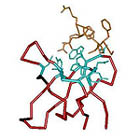
Diagram showing the positions of simplified residues (I, K, E, A, G) in red for FP2 in the wild type SH3 structure. Side chains of residues involved in ligand binding are displayed and residues where simplification was not attempted are in light blue. The peptide ligand is shown in orange. Residues which did not tolerate simplification are in black.